Frontal Muscle Anatomy
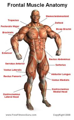
Major Muscles on the Front of the Body
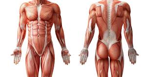
Our body has several layers of muscles. This list shows the outermost layer, called the superficial layer, of our major muscles. Muscles are usually work in pairs because although they can contract and shorten (flex), they are pulled by an opposite (antagonist) muscle to straighten out (extend) again. Sometimes the name of the muscle includes it’s function—such as extensor, flexor, adductor, abductor. The muscles on the front of the trunk help lift the arms and move the body forward and sideways. They also protect the organs in the abdomen.
To better understand muscles and how they work, it’s important to familiarize ourselves with the different planes of the body. In anatomy, there are three different planes in which our bodies move.
The frontal plane divides the body into a front and back. Movements occurring in the frontal plane include adduction and abduction. The saggital plane divides the body into a left and right side. This plane is where flexion and extension occurs. Finally, the transverse plane divides the body into a top and bottom. The movement that occurs in the transverse plane is rotation.
Contents
1 List of Major Anterior Muscles
1.23 Tensor fasciae lata (TFL)
List of Major Anterior Muscles
Major Muscles on the Front of the Body
The Coracobrachialis is the smallest of the three muscles that attach to the coracoid process of the scapula. (The other two muscles are pectoralis minor and biceps brachii.) It is situated at the upper and medial part of the arm. It is perforated by and innervated by the musculocutaneous nerve. The coracobrachialis draws the humerus forward (shoulder flexion) and towards the torso (shoulder adduction) at the glenohumeral joint.
Deltoid
The large triangular muscle that wraps around the shoulder and connects the scapula, clavicle and humerus. The deltoid muscle is referred to by its anterior, middle, and posterior fibers and is controlled by the axillary nerve.The shoulder muscle originates from the clavicle and scapula and inserting into the lateral part of the humorous. The front fibers help flex the arm, lateral fibers help abduct the arm and back fibers help extend the arm. Deltoid means triangular.
Extensor hallucis longus
The Extensor hallucis longus is a thin muscle, situated between the Tibialis anterior and the Extensor digitorum longus that extends the big toe, dorsiflexes the foot, and helps invert the foot. It arises from the anterior surface of the fibula; it also arises from the interosseous membrane to a similar extent. The anterior tibial vessels and deep peroneal nerve lie between it and the Tibialis anterior.
Extensor digitorum longus
This muscle arises from the lateral condyle of the tibia.Between it and the Tibialis anterior are the upper portions of the anterior tibial vessels and deep peroneal nerve. The muscle passes under the transverse and cruciate crural ligaments along with the Peronæus tertius, and divides into four slips, which run along the top of the foot and insert into the second and third phalanges of the four lesser toes.
Report this ad
The large muscle of the posterior part of the lower leg. It is the most superficial of the calf muscles. The gastrocnemius has 2 heads, one originating along the outside of the head and condyle of the femur and the other originating along the medial popliteal surface of the femur. Both heads attach to the back surface of the calcaneus, also called the heel bone the heel with the calcaneal tendon, also called the Achilles. When it contracts the gastrocnemius plantar flexes the ankle bending the foot downward, flexes the knee, and allows a person to stand on tip toes. It is innervated by the tibial nerve. It’s name means “belly of the leg.”
Gluteus medius
A muscle of the hip originating on the lateral surface of the ileum and inserted in the greater trochanter of the femur. It abducts and medially rotates the by and is controlled by the superior gluteal nerve.
Gracilis
A long slender muscle on the medial aspect of the thigh.
Iliopsoas
The compound iliacus and psoas magnus muscles.
Iliotibial band (ITB)
– a dense band of non-contractile tissue, called fascia, that covers the gluteal region and into this tensor fascia lata of an gluteus maximus are attached. Distallly the ITB inserts into the kneecap, tibia and fibula head.
Latissimus dorsi
A back muscle that pulls the arm down and back. It is responsible for extension,adduction, and (medial) internal rotation of the shoulder joint. It also helps in extension and lateral flexion of the lumbar spine. The name means “widest of the back.” This muscle supports the arm when it is moved above the head. If you push your arm hard against your side, you will feel this muscle tighten up.
Pectineus
Pectoralis major
A chest muscle that pulls the arm in towards the body. This is one of the internal rotator muscles that attach the humerus and internally rotate the arm. The pectoralis major originates along the clavicle, down the sternum, and across the ribs and inserts into the humerus. This muscle can contribute to excessive internal rotation of the arm or scapular abduction.
Peroneus longus
Fibularis longus muscle. A muscle along the outside of the leg that bends the foot out at the ankle. The fibularis longus originates from the head and upper lateral surface of the fibula, runs in a bony groove along the bottom of the foot to attach on the other side at the base of the first metatarsal and the neighboring medial cunieform bone, and acts to evert the foot; it is innervated by the superficial fibular nerve.
Rectus abdominis
Aalso known as the “six pack”, is a paired muscle running vertically on each side of the front wall of the abdomen. There are two parallel muscles, separated by a midline band of connective tissue called the linea alba. The rectus abdominis is an important postural muscle. It is responsible for pulling the rib cage toward the pelvis. The rectus abdominis helps when we exhale while breathing and forcefully exhaling. It also helps in keeping the internal organs intact and in creating pressure inside the abdomen, such as when exercising or lifting heavy weights, during forceful defecation or pushing during childbirth.
Rectus Femoris
a muscle of the anterior thigh originating on the iliac spine and upper margin of the acetabulum and inserted in the tibial tuberosity by way of the patellar ligament. It extends the leg, contributes to flexion of the thigh, and is controlled by the femoral nerve.
Sartorius
A long, ribbon-shaped muscle in the leg that flexes, abducts, laterally rotates the thigh, and flexes the lower leg. This muscle, the longest in the body, enables the crossing of the legs in the tailors’s position, the function for which it is named. It is strapped shaped and winds across the front of the thigh, from the hip to the inner side of the tibia. When it contracts it bends and rotates the thigh.
Serratus anterior
This muscle is divided into three named parts: serratus anterior superior, serratus anterior intermediate, serratus anterior inferior and runs from the front of the chest around the side to the scapula. The anterior serratus pulls the scapula outward which lifts the shoulder. It keeps the scapula in position close to the chest wall, abducts the scapula, and turns it upward to raise the point of the shoulder. If the scapula is fixed, the serratus anterior can elevate the ribs. The serratus anterior is controlled by the long thoracic nerve. Serratus means “saw-shaped” and describes this muscle’s jagged shape.
Sternocleidomastoid
is a paired muscle in the superficial layers of the front part of the neck. It tilts the head to its own side and rotates the head so the head faces the opposite side. It is also an accessory muscle of breathing out and raises the sternum.
Tensor fasciae lata (TFL)
originates on the anterior portion of the iliac crest and ASIS and inserts into the ITB. It flexes, medially rotates, and abducts the leg and can cause pelvic rotation problems.
Teres major muscle
is a muscle of the arm and one of six scapulohumeral muscles. It is not part of the rotator cuff. The teres major is a medial rotator and adductor of the humerus and assists the latissimus dorsi in drawing the previously raised humerus down and backward (extension, but not hyper extension). It also helps stabilize the humeral head in the glenoid cavity.
Tibialis anterior
An extensor muscle that straightens or lifts the foot. A muscle of the leg originating on the lateral condyle of the tibia and the interosseus membrane between the tibia and the fibula and inserted in the first cunieform and first metatarsal bones. It dorsi flexes and inverts the foot, supports the arch, and is controlled by the deep peroneal nerve.
Vastus lateralis
A muscle of the anterior thigh originating on the linea aspera and the greater trochanter of the femur and inserted in the tibial tuberosity by way of the patellar ligament. It extends the leg and is controlled by the femoral nerve.
Vastus medialis
a muscle of the anterior thigh originating on the linear aspera and inter-trochanteric line of the femur and inserted in the tibial tuberosity via the patellar ligament. It extends the leg and is controlled by the femoral nerve.
Glossary
ASIS
anterior superior iliac spine.
Distal
Farther from the midline. For instnace, the wrist is distal to the elbow joint – that is, the wrist is farther away from the midline than the elbow.
Proximal
Closer to the midline, that is – the elbow is distal to the wrist.
Adduction
Movements that bring limbs towards the midline. For instance, bringing your arm down to your from sideward extension is adduction.
Abduction
To abduct a limb or to move it away from the midline. Lifting the arms sidewards from resting position is an example of abduction.
Extension
Movements that straighten out a joint, for instance extending the knee involves straightening the knee joint.
Flexion
Shortening of a muscle. Flexion will often bend a limb, such as when flexing the bicep, thus bending the elbow.
Rotation
Movement that occurs in the horizontal plane. For instance, when the arms are at rest at a person’s sides, external and internal rotation will supinate or pronate the hands and forearms.
Insertion
The distal attachment of a muscle. The insertion is the segment that moves during muscle flexion. For instance, the bicep inserts along the radial tuberosity. When the bicep is flexed, the radius or forearm moves towards the upper arm.
Origin
The proximal attachment of the muscle, often considered the anchor of movement. For instance, the bicep originates from the scapula and shoulder. Thus any movement performed by the biceps will bring the insertion closer to the origin.
Innervation
Nerve supply of a muscle. Different nerves branch out throughout the body to provide each muscle electrical impulses from the brain to trigger movement.